Thrombosis
Deep vein thrombosisprevent & treat
A sudden swelling of an extremity with a feeling of tightness and heaviness, a new visibility of superficial veins or a bluish discolouration of an affected leg or arm - all of these can be Symptoms of deep vein thrombosis be. And it doesn't even have to cause severe pain at the beginning. Nevertheless, if you suffer from the symptoms mentioned, you should quickly seek medical advice. Specialist treatment because an initial thrombosis can develop into a thrombosis dangerous develop pulmonary embolism.
Diagnosis & Therapy
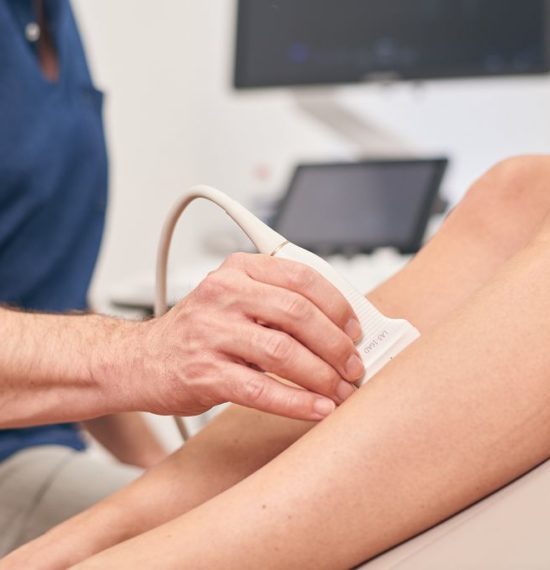
Rapid diagnostics with ultrasound
Thrombosis is a blockage of a deep vein in the leg or arm caused by a blood clot. However, the pelvic veins or the inferior vena cava can also be affected by thrombosis. The lack of blood flowing back to the heart then causes the recognisable swelling of the affected extremity.
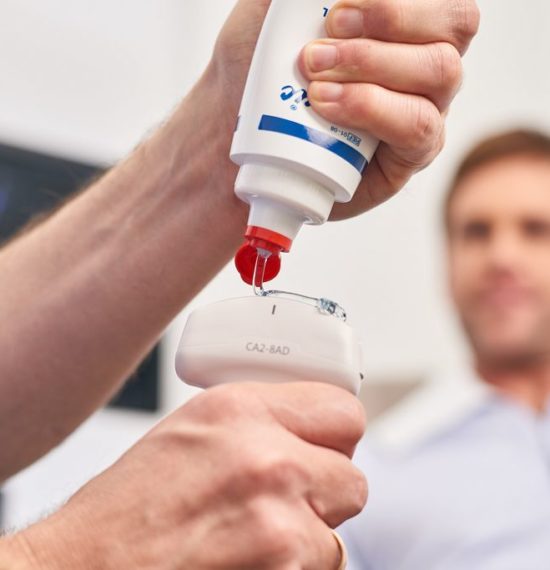
Ultrasound examinations are used for diagnostics, with which the blood vessels in the legs can be analysed. visualised can be realised. It is painless and completely harmless. If the vein can be easily compressed with the transducer, there is no thrombosis. However, if the vein cannot be compressed because a clot inside it is causing it to harden, there is almost always a thrombosis.
How is a fresh deep vein thrombosis treated?
Nowadays, thanks to modern blood-thinning medication, deep vein thrombosis outpatient treatment become.
To prevent the further spread of thrombosis prevent and swelling of the extremity to counteract, In the acute stage, a compression bandage is applied and a blood-thinning medication - usually in the form of a tablet - is prescribed. Once the swelling of the extremity has subsided, the patient is given a compression stocking for several months. In the area of the pelvic vein, the clot can sometimes be dissolved using special medication (known as lysis) or the Surgical removal with a catheter. However, this is a case-by-case decision and depends on the individual patient.

Post-thrombotic syndrome
As possible Late effect deep vein thrombosis can lead to Damage to other vein sections which then occur in the so-called Post-thrombotic syndrome open up. This means that the deep veins can no longer fulfil their function of transporting blood from the legs towards the heart and the blood backs up into the legs. This then leads to persistent leg swelling, new varicose veins and leg ulcers. The therapy usually consists of lifelong wearing of compression stockings. It is therefore crucial to wear compression stockings after a deep vein thrombosis in regular medical check-ups in order to prevent post-thrombotic syndrome. With a special and painless Investigation, the Vein occlusion plethysmography (VVP), your vein function can be checked after a thrombosis has occurred.
Frequently asked questions on the topic of "thrombosis"
A thrombosis is a blood clot in a vein, usually in the leg or arm. A distinction must be made between superficial and deep vein thrombosis. In the case of a superficial vein thrombosis, which usually occurs in a visible varicose vein in the leg, there is a string-like and hard thickening of the corresponding vein with reddening and significant pain. In the case of deep vein thrombosis, there is an obstruction to the outflow of blood from the affected limb, resulting in significant swelling of the leg or arm with a feeling of tightness. Exceptions to this are muscle vein thrombosis or isolated lower leg vein thrombosis, which usually only lead to pain and not to swelling. In any case, if a thrombosis occurs, it must be clarified in detail using ultrasound in order to determine its true extent, as it is only visible from the outside for a short distance, but can be significantly more extensive in the depth of the leg.
In my practice, the type of thrombosis (superficial, deep, muscular vein) is first clarified with a physical examination and then with ultrasound. Depending on the location and extent of the thrombosis, blood is then thinned for between 6 weeks and 6 months or, if necessary, for life - depending on the severity of the thrombosis. Wearing a compression stocking on the leg or arm helps to reduce swelling and discomfort and appears to speed up the breakdown of the blood clot. Hospitalisation for thrombosis is no longer necessary. I initiate the relevant investigations into the surrounding area to clarify the cause of the thrombosis, which are usually carried out by the family doctor. A first check-up usually follows after approx. 3 weeks.
If a thrombosis is not recognised in time and blood thinning (anticoagulation) is not initiated, this can lead to the thrombosis growing and possibly to an additional pulmonary embolism, which can also be dangerous.
Risk factors for thrombosis are often prolonged immobility (travelling for long periods without getting up or lying down during infections) or the combination of the pill and smoking. Sometimes there is also a genetic predisposition to thrombosis, but this can only be determined by a large coagulation test. In rare cases, tumours also lead to thrombosis, so a tumour must also be ruled out in the case of unexplained thrombosis.
The body often breaks down a thrombosis over the course of weeks to a few months. However, it is also possible for a thrombosis to remain completely or partially intact. However, this does not necessarily mean that it will be a problem for the rest of your life. Rather, the prognosis (persistent swelling of the leg or arm) depends on whether the remaining or broken down thrombosis has led to vein damage or to a drainage disorder from the affected vein. This can also be determined during the course of the procedure using ultrasound.
Impressions of the practice